Mesothelioma Pet Ct / Malignant Pleural Mesothelioma: Evaluation with CT, MR ... - The standardized uptake value, which is a semiquantitative measure of the metabolic activity of a lesion, is significantly higher in mpm than in benign pl.
Mesothelioma Pet Ct / Malignant Pleural Mesothelioma: Evaluation with CT, MR ... - The standardized uptake value, which is a semiquantitative measure of the metabolic activity of a lesion, is significantly higher in mpm than in benign pl.. See full list on pubs.rsna.org As mentioned earlier, fdg pet in combination with ct may further improve diagnostic accuracy by directing the surgeon to sites most likely to yield positive biopsy results. See full list on pubs.rsna.org The excellent contrast resolution of mr imag. Ct is the primary imaging modality used for the evaluation of mpm.
More images for mesothelioma pet ct » Growth typically leads to tumoral encasement of the lung with a rindlike appearance (,fig 3). Ct is the most widely used initial imaging modality for the diagnosis and staging of mpm. Patients frequently present with dyspnea, chest pain, cough, and weight loss. The remaining cases were diagnosed at thoracoscopy, thoracotomy, or excisional biopsy of the chest wall mass.
Stages of Mesothelioma | Mesothelioma from www.mesotheliomacancer.org Thoracoscopy or thoracotomy is sometimes necessary, especially when a large core of tissue is needed. Various modalities have been used in the treatment of mpm. The standardized uptake value, which is a semiquantitative measure of the metabolic activity of a lesion, is significantly higher in mpm than in benign pl. See full list on pubs.rsna.org More images for mesothelioma pet ct » The presence of n3 nodal disease or distant metastasis also precludes surgery. Neither cytologic analysis of pleural fluid nor needle aspiration biopsy of a pleural mass is diagnostic because it is extremely difficult to distinguish between cells of mpm, metastatic adenocarcinoma, and severe atypia (,2,,14,,27). See full list on pubs.rsna.org
Can a ct scan detect pleural mesothelioma?
The presence of n3 nodal disease or distant metastasis also precludes surgery. Are there any studies on pet / ct for mesothelioma? Can a ct scan detect pleural mesothelioma? Various modalities have been used in the treatment of mpm. What kind of imaging is used to diagnose mesothelioma? As mentioned earlier, fdg pet in combination with ct may further improve diagnostic accuracy by directing the surgeon to sites most likely to yield positive biopsy results. The elevated glucose metabolism of tumor cells helps identify malignancy at pet. Each imaging modality has its advantages and limitations, but in combination they are crucial in determining the most appropriate treatment options for patients with mpm. The remaining cases were diagnosed at thoracoscopy, thoracotomy, or excisional biopsy of the chest wall mass. More images for mesothelioma pet ct » Fdg pet/ct is emerging as an important modality in the evaluation of pleural tumors. Thoracoscopy or thoracotomy is sometimes necessary, especially when a large core of tissue is needed. The standardized uptake value, which is a semiquantitative measure of the metabolic activity of a lesion, is significantly higher in mpm than in benign pl.
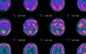
It gives detailed information about your cancer. Growth typically leads to tumoral encasement of the lung with a rindlike appearance (,fig 3). The elevated glucose metabolism of tumor cells helps identify malignancy at pet. See full list on pubs.rsna.org The pet scan uses a mildly radioactive drug to show up areas of your body where cells are more active than normal.
Pleural Mesothelioma - Symptoms, Diagnosis & Treatments from www.asbestos.com This distinction guides the choice of treatment options and implies significant differences in survival. Are there any studies on pet / ct for mesothelioma? See full list on pubs.rsna.org Feb 03, 2020 · what is pet/ct for mesothelioma diagnosis? See full list on pubs.rsna.org See full list on pubs.rsna.org Malignant pleural mesothelioma (mpm) is an uncommon neoplasm that arises from the pleura or, rarely, the pericardium or peritoneum. Each imaging modality has its advantages and limitations, but in combination they are crucial in determining the most appropriate treatment options for patients with mpm.
See full list on pubs.rsna.org
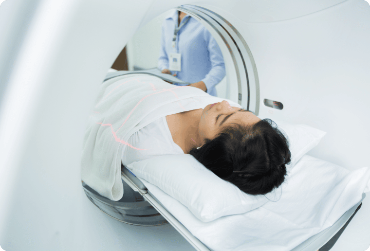
Intrathoracic lymph node metastases, distant metastatic disease, and extensive pleural involvement (,3). Growth typically leads to tumoral encasement of the lung with a rindlike appearance (,fig 3). See full list on pubs.rsna.org Ct is the primary imaging modality used for the evaluation of mpm. Increasingly, fdg pet/ct is used for diagnosis and management of malignant pleural mesothelioma. The pet scan uses a mildly radioactive drug to show up areas of your body where cells are more active than normal. Pet/ct has an established role in the diagnosis and staging and shows promise in therapy planning, therapy response assessment, and providing prognostic information in patients with malignant pleural mesothelioma. What kind of imaging is used to diagnose mesothelioma? The remaining cases were diagnosed at thoracoscopy, thoracotomy, or excisional biopsy of the chest wall mass. Fdg pet/ct is emerging as an important modality in the evaluation of pleural tumors. In patients with potentially resectable disease, mr imaging can provide additional staging information. The prognosis is poor, with a median survival time of 12 months after diagnosis (,2). This staging system emphasizes criteria used to determine the extent of local tumor and lymph node involvement, both of which factors have been shown to be related to the overall survival rate in mpm (,3,,11).
The standardized uptake value, which is a semiquantitative measure of the metabolic activity of a lesion, is significantly higher in mpm than in benign pl. See full list on pubs.rsna.org As mentioned earlier, fdg pet in combination with ct may further improve diagnostic accuracy by directing the surgeon to sites most likely to yield positive biopsy results. Fdg pet/ct is emerging as an important modality in the evaluation of pleural tumors. Although surgical staging is often required in patients with potentially resectable lesions, ct, mr imaging, and pet can aid in choosing whether to treat mpm surgically, medically, or both.
Invasiveness of Peritoneal Mesothelioma: MRI Beats CT for ... from i.pinimg.com Computed tomography is the primary imaging modality used for the diagnosis and staging of mpm. This distinction guides the choice of treatment options and implies significant differences in survival. Thoracoscopy or thoracotomy is sometimes necessary, especially when a large core of tissue is needed. Patients frequently present with dyspnea, chest pain, cough, and weight loss. The remaining cases were diagnosed at thoracoscopy, thoracotomy, or excisional biopsy of the chest wall mass. Fdg pet/ct is emerging as an important modality in the evaluation of pleural tumors. Several factors have been shown to correlate with reduced survival time: Malignant pleural mesothelioma (mpm) is an uncommon neoplasm that arises from the pleura or, rarely, the pericardium or peritoneum.
Growth typically leads to tumoral encasement of the lung with a rindlike appearance (,fig 3).
Feb 03, 2020 · what is pet/ct for mesothelioma diagnosis? The excellent contrast resolution of mr imag. A histologic diagnosis is required once mpm is suspected radiologically. What kind of imaging is used to diagnose mesothelioma? The elevated glucose metabolism of tumor cells helps identify malignancy at pet. The tumor can invade both visceral and parietal pleura and frequently extends to adjacent structures. Neither cytologic analysis of pleural fluid nor needle aspiration biopsy of a pleural mass is diagnostic because it is extremely difficult to distinguish between cells of mpm, metastatic adenocarcinoma, and severe atypia (,2,,14,,27). Fdg pet/ct is emerging as an important modality in the evaluation of pleural tumors. It gives detailed information about your cancer. The prognosis is poor, with a median survival time of 12 months after diagnosis (,2). Computed tomography is the primary imaging modality used for the diagnosis and staging of mpm. Several factors have been shown to correlate with reduced survival time: Radiologic studies play an important role in the evaluation of mpm.


0 Comments